Pathology
Tumors of the head and neck, also called cervicofacial or upper aerodigestive tract cancers, include several neoplasms that develop in areas such as the mouth, tongue, gums, pharynx, larynx, nose, sinuses, and salivary glands and may involve the development of metastases to the lymph nodes in the neck. The most frequently affected site is the larynx, followed by the oral cavity and pharynx
These tumors affect a part of our body that is fundamental in everyday life and can result in a major alteration on function and aesthetics.Therefore, the treatment path requires a multidisciplinary approach that also integrates rehabilitation and psychological support.
Thecourse of the disease depends greatly on the site of the tumor, its stage at the time of diagnosis, and the patient’s general condition. In many cases, especially when detected early and treated in specialized centers, the chances of cure and control of these tumors are comforting.
Types
Most of these neoplasms are spinocellular carcinomas, which originate from squamous cells lining mucosal surfaces;
Less common, but still relevant, are melanomas, lymphomas, sarcomas, and salivary gland tumors.
The numbers in Italy
According to the AIRTUM registry (Italian Association of Cancer Registries), head and neck cancers in Italy in 2022 registered about 9,750 new diagnoses (7,050 men and 2,700 women).
Symptoms
Initial symptoms are often mild and nonspecific, making early diagnosis difficult. Among the most common signs:
- Pain in the tongue and gums;
- Persistent sore throat;
- Sense of constriction in the throat or difficulty swallowing;
- Dysphonia, as a persistent voice alteration
- difficulty breathing;
- nasal obstruction;
- Relapsing epistaxis;
- Ear pain (otalgia);
- Occurrence of swelling in the face or neck.
Risk factors
Known risk factors are:
- tobacco and alcohol, which are the main risk factors;
- Injuries and continuous small microtrauma from altered prostheses and poorly cared for teeth can lead to the development of tumors in the tongue;
- cancers of the nasopharyngeal tract are often associated with infection caused by the Epstein-Barr virus (EBV);
- cancers of the oropharynx may be related to human papilloma virus (HPV) infection.
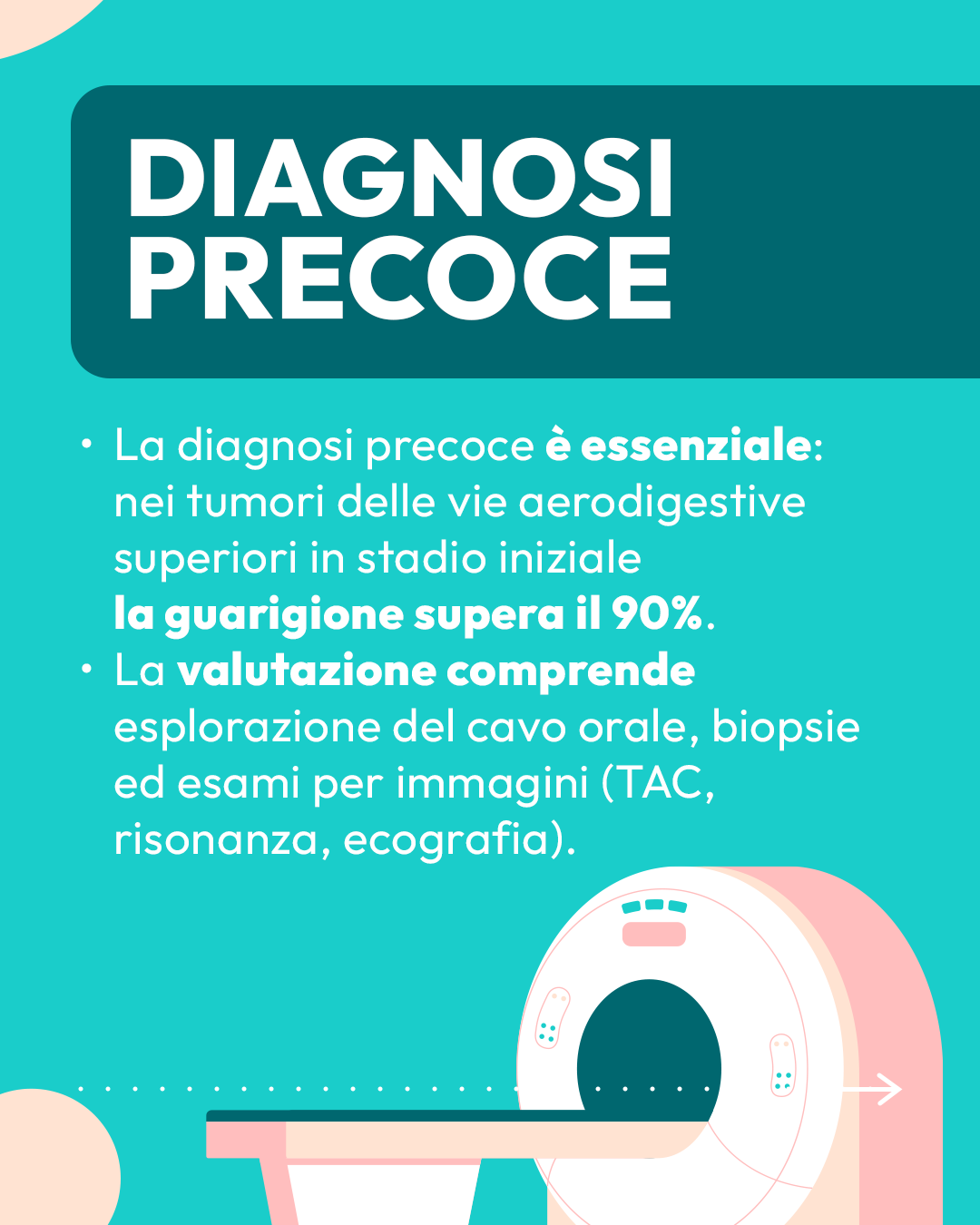
Diagnosis and examination
For proper diagnosis of the disease and its extent, a thorough patient history, objective examination supplemented by instrumental investigations, and radiological imaging examinations are of paramount importance.
Instrumental examinations
Direct fiberoptic laryngoscopy: consists of direct visualization of the nasal passages, pharynx and larynx. It is performed by passing a fibroscope (a small-diameter flexible tube consisting of an optic and a camera) through the nose to visualize the pharynx and larynx at its different levels. It is an examination well tolerated by the patient; it allows excellent visualization of the upper airway and is aimed at identifying direct or indirect signs of disease.
The instrumental equipment of the Candiolo Institute’s outpatient clinics also allows the performance of fibroscopy with NBI light, which represents a diagnostic innovation today, as it visualizes mucosal areas suspicious for precancerosis ,through visual analysis of aberrant submucosal vasculature.
Diagnostic imaging examinations
Ultrasound of the neck
This is a first-level, noninvasive examination. It allows the evaluation of neck lymph nodes. Lymph nodes can be sites of metastasis of head and neck cancer. Ultrasound can be completed with a needle biopsy Of a lymph node in the neck if suspicious. Using a special needle, cells from the lymph node are to be harvested and sent to the Anatomopathologist for analysis (cytological examination).
Chest X-ray
This is a first-level, noninvasive examination that allows a photograph of the chest Of the patient, with special attention to the lungs. This examination is very important both in the preoperative, in case the patient should undergo surgery, but also to assess the possible presence of lung metastases of the head-neck tumor, or the simultaneous presence of a lung tumor. In fact, smoking is the most important cause of lung cancer as well.
CT, MRI, PET
The Tcomputed tomography (CT), the Magnetic Resonance Imaging (MRI) and the Positron Emission Tomography (PET) are second-level examinations that are essential in the evaluation of head and neck cancer. They provide Information about the location and depthextent of the tumor and identify the presence of metastasis to lymph nodes in the neck or other locations in the body. They are often essential for planning surgery or for indication for radiation or chemotherapy treatments.
Therapies
After the diagnosis is confirmed, specialists in the multidisciplinary team evaluate a number of factors to plan an individualized course of treatment for the patient. In addition to the type of tumor, its size, and whether it has spread to other parts of the body, the patient’s age, general health status, and medical history are also considered. The treatment plan is then discussed with the patient, proposing alternative choices in case of equivalent effectiveness.
Depending on the stage of the tumor, different treatment strategies are indicated including surgery, chemotherapy, radiation therapy, and molecular targeted therapies.
Surgery
In head and neck cancers, the treatment of first choice is often surgery, which today can also provide excellent results with minimally invasive approaches and targeted excisions. These surgeries, performed without skin incisions, allow for a short hospitalization, reduced convalescence, and rapid resumption of normal activities. In particular, transoral CO₂ laser surgery is an effective option for treating early-stage cancers of the mouth, pharynx, and especially the larynx.
In more advanced cases, however, surgery often involvesremoval of the tumor combined with removal of neck lymph nodes potentially involved in the disease.
The Candiolo Institute is equipped with state-of-the-art technologies that allow even extensive surgeries to be performed while preserving the functionality of surrounding organs and nerve structures. One example is the routine use of intraoperative neuromonitoring in major salivary gland and thyroid surgery, with the goal of minimizing the risk of iatrogenic nerve damage and ensuring that the patient receives the highest level of treatment.
In surgeries for advanced tumors, in addition to complete removal of the disease, it is essential to safeguard the cosmetic and functional appearance of the affected district. This is why we resort to reconstructive surgery, which improves the chances of recovery of swallowing and breathing. At the Candiolo Institute, this technique makes it possible to restore organ function by transplanting tissue taken from other parts of the body. Through complex but highly specialized surgeries, portions of the tongue, oropharynx, or palate can be reconstructed, achieving outstanding functional and aesthetic results and preserving a good quality of life.
After surgery, definitiveanatomopathologic analysisis performed, assessing the tumor for size, degree of aggressiveness, possible extension to nearby structures and lymph nodes, with special attention to resection margins. Based on these data, complementary treatment, such as radiotherapy combined or not with chemotherapy, may be indicated.
Chemotherapy
Chemotherapy is a drug treatment that aims to eliminate cancer cells by exploiting their faster rate of reproduction than healthy cells. Because it acts by interfering with cell replication mechanisms, it can also affect healthy cells, causing side effects that, in most cases, disappear once the treatment is over.
Before starting, the oncologist provides all the information about the drugs to be used and useful behaviors to alleviate any side effects.
In head and neck cancer–particularly in oropharyngeal or nasopharyngeal forms related to viral infection, and in advanced laryngeal cancers–the combination of radiotherapy and chemotherapy can lead to cure in a high percentage of cases. This strategy preserves pharyngeal and laryngeal function even in locally extensive tumors in which surgery is not indicated.
Chemotherapy can also be used:
-
As the exclusive treatment in advanced or metastatic head and neck cancers;
-
in combination with radiation therapy after surgery if histologic examination shows particularly aggressive forms.
Administration is usually intravenous, on an outpatient basis. The duration of each administration varies from a few minutes to several hours, depending on the drugs used.
The treatment is carried out in cycles: each cycle lasts a few days and is followed by a rest period of several weeks. The total number of cycles depends on the type of tumor and the patient’s individual response to therapy.
Biological therapies
Biologic therapies, also knownas molecular targeted therapies(target therapies), are targeted treatments that selectively act on specific molecular targets-such as receptors, growth factors or enzymes-that are found primarily in cancer cells.
These targets are involved in fundamental disease processes, such as:
-
The uncontrolled growth and spread of cancer cells;
-
resistance to traditional therapies;
-
The formation of new blood vessels that nourish the tumor(angiogenesis).
One of the most important targets is EGFR(Epidermal Growth Factor Receptor), a receptor on the surface of cancer cells that binds to epidermal growth factor. Activation of EGFR stimulates the RAS gene, which is responsible for numerous proliferation and metastatic processes.
Targeted drugs, such as Cetuximab, a laboratory-produced monoclonal antibody that mimics the action of the immune system and binds to EGFR, inhibiting its activity, are used to block this growth pathway.
Immunotherapy
Immunotherapy includes drugs that are not directed against tumor cells but work by activating the immune system response blocked by the tumor.
For these cancers, researchers are studying drugs that can take the brakes off the immune system, thus making it effective again in recognizing and eliminating cancer cells.
These are still experimental therapies, applied for now in certain types of metastatic cancer, but they seem to have a promising future.
Radiotherapy
Radiation therapy uses high-energy radiation to destroy cancer cells.
It is an outpatient treatment, which does not require hospitalization, administered at the Division of Radiotherapy in consecutive daily sessions from Monday to Friday, organized in cycles varying in duration from one to several weeks.
Because tumors in the head and neck region develop near particularly delicate organs-such as the eyeball, nervous system, or trachea-it is essential to have state-of-the-art equipment that allows for intensity-modulated radiation therapy. This technique allows the photon beam to be focused on the tumor target, sparing nearby healthy structures as much as possible and thus reducing side effects. One example is Tomotherapy, which combines pinpoint precision with treatment options on complex areas.
Thanks to modern techniques, serious complications of radiation therapy are now reduced to 1-2%. Frequent late side effects include some dryness of irradiated mucous membranes and reduced sense of taste.
In head and neck cancer, radiation therapy can be:
-
associated with chemotherapy as first-choice therapy, particularly in virus-related forms and in some advanced laryngeal cancers;
-
indicated after surgery (adjuvant therapy) when histologic examination shows particularly aggressive forms, to reduce the risk of recurrence;
-
used as an alternative to minimally invasive transoral surgery in early-stage cancers of the larynx, soft palate, and oropharynx;
-
used for palliative purposes, to reduce pain or relieve symptoms due to infiltration of head and neck organs.
Ongoing support
At our institute, we ensure constant support before, during and after treatment to accompany each patient throughout the entire course of treatment and recovery.
Psychological support
The impact of cancer in a person’s life also affects the psychological sphere: falling ill with cancer is in fact always a traumatic event that affects all dimensions of the person and can generate anxiety, fear, anger, depression.
At the Candiolo Institute, alongside cutting-edge therapies, the treatment and care pathway always includes a qualified psycho-oncological support that helps the patient cope positively not only with treatment but also with the delicate phase of physical and psychological recovery.
It is also possible to participate in support groups psychological to compare with other people who have gone through or are going through the same experience.
Direct line to specialists
To ensure timely and direct support and receive timely answers to concerns and questions, a dedicated support service is in place at the Candiolo Institute for all patients.
From Monday to Friday, from 8 a.m. to 5 p.m., you can contact the secretariat of the oncology day hospital at 011.993.3775, reporting the need for urgent consultation.
The patient will be quickly put in touch with his or her medical specialist, to receive clear answers and immediate support.
Continuing and palliative care
The cancer patient is a person with complex needs that requires multidisciplinary support not only for the cancer disease, but also for all related issues.
At the Candiolo Institute, patients who need or require it have access to specialists in different areas to receive nutritional support, physical therapy, pain therapy and management of other associated conditions.
Social work
The Social Service Department of the Candiolo Institute conducts information and orientation interviews to patients and their families on how to access services in the area and how to obtain welfare and social security benefits provided by law (disability, benefits for aids and prostheses, work leave, etc.).
The service operates on Wednesdays and Fridays from 9 a.m. to 1 p.m. (phone: 011 9933059).
Follow up
With the conclusion of the course of treatment, the follow-up period begins, during which, through medical examinations and a series of tests, we monitor:
-
The side effects of the therapies carried out;
-
The effectiveness of treatments;
-
The functional recovery of the patient.
Follow-up examinations are especially crucial to intercept any recurrence early, so that prompt action can be taken with the most appropriate therapy. For the patient, they also represent an important opportunity for discussion and dialogue with their medical specialist.
It is the same specialist physician who schedules follow-up visits, during which the patient’s general health condition is assessed and the required test reports are analyzed.
The checks follow scheduled intervals for the duration of five years. They have a shorter cadence at first, every three months, and then gradually thin out over time to twice a year. The frequency and type of examinations provided depend on the stage of the tumor and the treatments performed.
In head and neck cancer, follow-up usually involves:
-
Medical examination with fibrolaryngoscopy: every 3 months in the first 2 years, then every 6 months in the 4th and 5th years;
-
Instrumental examinations: CT scan of the neck and chest, MRI of the facial massif and neck, or CT/PET scan at 3-6 months after the end of treatment, then every 6 months thereafter for the first 2 years and every 6-12 months until the 5th year.
These tests can also be requested at any time if a recurrence of illness is suspected during follow-up visits.
Interdisciplinary Group
Every cancer requires, in all phases of disease management, a multidisciplinary approach that at the Candiolo Institute is guaranteed by a team of different specialists, belonging to the various clinical and surgical departments of the Institute: this team is called GIC (Interdisciplinary Care Group). The GIC ensures that each patient is taken care of throughout the diagnostic-therapeutic process, including prescribing and booking examinations and communicating with the patient and his or her family members. The GIC defines and shares a personalized care pathway for each patient, based not only on the type and stage of the tumor, but also on the patient’s own characteristics. The goal is to ensure that he or she has the best outcome both oncologically and functionally and the maintenance of a good quality of life.The Group also works closely with researchers at the Institute to ensure that patients have rapid access to the latest research-produced innovations in screening, diagnosis and treatment.
Clinical divisions
The head and neck cancer diagnostic and therapeutic pathway at Candiolo involves several clinical divisions, including:
- Otolaryngology
- Reconstructive plastic surgery
- Anesthesia and resuscitation
- Medical oncology
- Nuclear medicine
- Radiotherapy
- Radiodiagnostics
- Pathologic anatomy
Clinical studies
At the Candiolo Institute for Head and Neck Cancer, scientific research is particularly advanced.
With patients’ consent, small samples of the tumor are taken and stored in the laboratory so that the cells remain alive. These samples are transformed into organoids, which are small versions of the tumor, like “mini-tumors” in a test tube. Organoids allow researchers to study the characteristics and evolution of the tumor and to understand how it responds to drugs.
The mini-tumor biobank serves two main purposes:
- Test the drugs already used to understand which ones work best and why they sometimes do not work;
- try new drugs or drugs already known for other diseases to find out if they can also help against these cancers.
There are no other cancer centers in Italy with such a large number of organoids dedicated to head and neck cancers on which DNA sequencing, which identifies the genetic profile of cancer cells, has also been performed.
Thanks to this research, the Institute can test new treatments in the laboratory before using them on patients.
Although the final results are not yet available, the preliminary data are very promising.
Why choose us
At Candiolo IRCCS Institute, every head and neck cancer patient is followed according to highly specialized standards, thanks to the synergistic work of a dedicated Interdisciplinary Care Group (ICG).
Clinical experience and tailored approach
Due to the high number of cases treated each year, the Candiolo Institute is a national reference for taking care of esophageal cancer. Our experience enables us to deal with even the most complex situations, always with a personalized approach built on the clinical and personal profile of each patient.
Imaging technologies and advanced diagnostics
Establishing the treatment plan always starts with an accurate and timely diagnosis. Patients have access to state-of-the-art imaging technologies that allow accurate assessment of the extent of the disease.
In addition, the Institute offers advanced and sophisticated laboratory investigations, including molecular and genomic analyses, which are critical for identifying biological features of cancer and guiding therapeutic decisions.
Minimally invasive surgical techniques and multidisciplinarity
When indicated, surgery is performed with minimally invasive techniques (laparoscopic or thoracoscopic), which reduce operative trauma, promote faster recovery, and improve postoperative quality of life. Every treatment choice is defined within the GIC, ensuring a consistent and integrated approach.
Clinical research and access to trials
As an IRCCS, the Candiolo Institute combines clinical practice with a strong vocation for scientific research. Patients can be evaluated for inclusion in active clinical trials, which represent a real chance to access innovative therapies not yet available in standard practice. Collaboration between care and research is a distinctive value that translates into concrete opportunities for the patient.
Care and support every step of the way
The Interdisciplinary Care Group takes care of the person at every stage: from diagnosis to treatment to follow-up, with attention to nutritional support, psychological health, and reintegration into daily life. The organization of checkups, visits and treatment is designed to ensure continuity and serenity, always valuing the human dimension of care.